From 11:00 pm to 12:00 pm EST ( 8:00 pm to 9:00 pm PST ) on January 6th, the website will be under maintenance. We are sorry for the inconvenience. Please arrange your schedule properly.
Chlorhydrate de doxorubicine (chlorhydrate d'hydroxydaunorubicine), un antibiotique anthracycline cytotoxique, est un agent de chimiothérapie anticancéreuse. Chlorhydrate de doxorubicine inhibe la topoisomérase, arrêtant ainsi la réplication de l'ADN. Chlorhydrate de doxorubicine réduit la phosphorylation basale de AMPK et de son cible en aval acétyl-CoA carboxylase. Chlorhydrate de doxorubicine induit l'apoptose et l'autophagie.
Doxorubicin hydrochloride (Hydroxydaunorubicin hydrochloride), ein zytotoxisches Anthracyclin-Antibiotikum, ist ein Chemotherapeutikum gegen Krebs. Doxorubicinhydrochlorid hemmt die topoisomerase II und stoppt so die DNA-Replikation. Doxorubicin-Hydrochlorid reduziert die basale Phosphorylierung von AMPK und seiner nachgeschalteten Ziel-Acetyl-CoA-Carboxylase. Doxorubicinhydrochlorid induziert apoptosis und autophagy.
Doxorubicin (Hydroxydaunorubicin) hydrochloride, a cytotoxic anthracycline antibiotic, is an anti-cancer chemotherapy agent. Doxorubicin hydrochloride is a potent human DNA topoisomerase I and topoisomerase II inhibitor with IC50s of 0.8 μM and 2.67 μM, respectively. Doxorubicin hydrochloride reduces basal phosphorylation of AMPK and its downstream target acetyl-CoA carboxylase. Doxorubicin hydrochloride induces apoptosis and autophagy.
For research use only. We do not sell to patients.
Doxorubicin or CPT-11 significantly promoted KRT8 expression in chordoma cells in vitro. Representative images of immunofluorescence staining of KRT8 of CM318 and UCH1 cell line.
Doxorubicin or CPT-11 significantly promoted KRT8 expression in chordoma cells in vitro. Western blotting analysis and quantification of KRT8 protein expression (normalized to GAPDH expression).
Effects of YWPC (50 mg/L for 24 h) on DOX (5 μM for 24 h)-induces dissipation of mitochondrial membrane potential measured in H9C2 cells loaded with JC-1 using fluorescence microscopy.
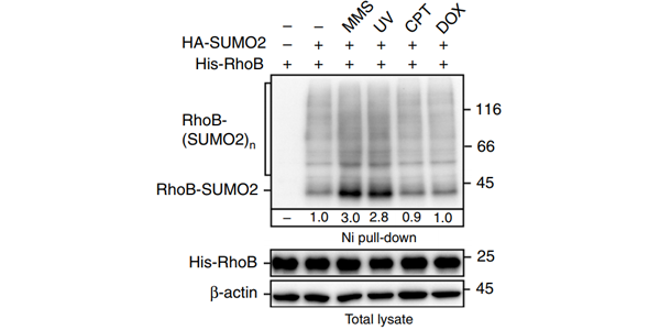
One hour after treated with UV or 2 h after treated with MMS, Camptothecin (CPT) (10 μM), or Doxorubicin (DOX; 0.5 μM), HEK293T cells expressing HA-SUMO2 and His-RhoB are subjected to sumoylation assay to detect the SUMO2 conjugation of RhoB.
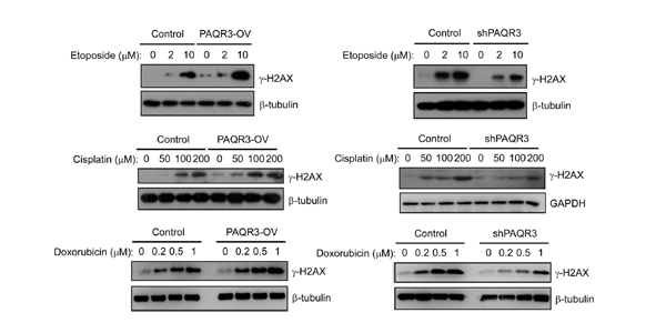
PAQR3 affects DNA damage repair. AGS cells are treated with different doses of VP-16 (for 24 h), CDDP (for 24 h) and Doxorubicin (for 10 h) as indicated, followed by immunoblotting with the antibodies.
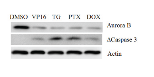
U937 cells are treated with Thapsigargin (TG, 1 μM), NSC 125973 (PTX, 5nM) as well as Doxorubincin (DOX, 1 μM) for 12 hours and the indicated proteins are detected by Western blot.
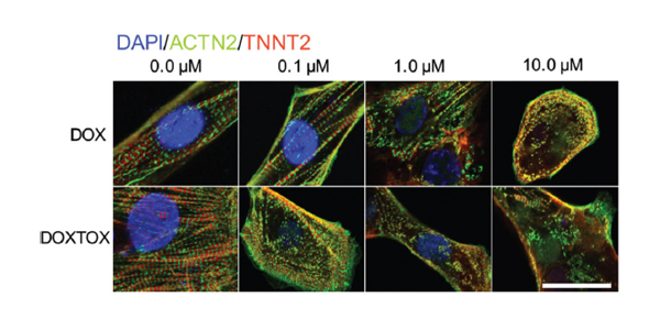
Immunofluorescent staining for α-actinin (ACTN2) and cardiac troponin T (TNNT2) to demonstrate sarcomeric organization in hiPSC–CMs derived from patients who do not experience Doxorubicin–induced cardiotoxicity (DOX) versus those who do experience Doxorubicin-induced cardiotoxicity (DOXTOX) after 24 h treatment with Doxorubicin. Scale bar, 20 µm.
Doxorubicin (Hydroxydaunorubicin) hydrochloride, a cytotoxic anthracycline antibiotic, is an anti-cancer chemotherapy agent. Doxorubicin hydrochloride is a potent human DNA topoisomerase I and topoisomerase II inhibitor with IC50s of 0.8 μM and 2.67 μM, respectively. Doxorubicin hydrochloride reduces basal phosphorylation of AMPK and its downstream target acetyl-CoA carboxylase. Doxorubicin hydrochloride induces apoptosis and autophagy[1][2][3].
Doxorubicin hydrochloride (1-8 μM; 24 and 48 hours) decreases the viability of MCF-10F, MCF-7 and MDA-MB-231 cells in a time- and dose-dependent manner[4].
Doxorubicin hydrochloride (1 μM; 3 and 24 hours) results in Hct-116 human colon carcinoma cells reduction in G0/G1 phase and accumulation in G2 phase[5].
Doxorubicin hydrochloride (1 μM for MCF-10F and MDA-MB-231 cells, 4 μM for MCF-7 cells; 48 hours) induces apoptosis by upregulating Bax, caspase-8 and caspase-3 and downregulation of Bcl-2 protein expression[4].
Doxorubicin can label neuron cells, and it is bright red under Rhodamine filter bag, and light red-orange under catecholamine filter bag[7].
Doxorubicin (5 μM; 10-30 min) can be accumulated in B16-F10 melanoma cell line CRL-6475 in a time-dependent manner, and can be detected by green or red fluorescence (green fluorescence has higher detection sensitivity) with a maximum excitation wavelength (λex) and a maximum emission wavelength (λem) of 470 nm and 560 nm, respectively[8]. Note:Doxorubicin hydrochloride is suitable for neutral or weakly acidic solvents; PBS (PH 7.4) is not recommended for dissolution, which may affect the dissolution effect.
MedChemExpress (MCE) has not independently confirmed the accuracy of these methods. They are for reference only.
Breast cancer cell lines MCF-10F, MCF-7 and MDA-MB-231
Concentration:
1 μM for MCF-10F and MDA-MB-231 cells, 4 μM for MCF-7 cells
Incubation Time:
48 hours
Result:
Bax protein expression was upregulated in MCF-10F and MDA-MB-231 cell lines but MCF-7 cells did not show any significant increase.
Caspase-8 gene expression was upregulated in MCF-10F, but it was downregulated in MCF-7 and MDA-MB-231 cells.
In Vivo
Doxorubicin hydrochloride (1-8 μM; 24 and 48 hours) decreases the viability of MCF-10F, MCF-7 and MDA-MB-231 cells in a time- and dose-dependent manner[4].
Doxorubicin hydrochloride (1 μM; 3 and 24 hours) results in Hct-116 human colon carcinoma cells reduction in G0/G1 phase and accumulation in G2 phase[5].
Doxorubicin hydrochloride (1 μM for MCF-10F and MDA-MB-231 cells, 4 μM for MCF-7 cells; 48 hours) induces apoptosis by upregulating Bax, caspase-8 and caspase-3 and downregulation of Bcl-2 protein expression[4].
Doxorubicin can label neuron cells, and it is bright red under Rhodamine filter bag, and light red-orange under catecholamine filter bag[7].
Doxorubicin (5 μM; 10-30 min) can be accumulated in B16-F10 melanoma cell line CRL-6475 in a time-dependent manner, and can be detected by green or red fluorescence (green fluorescence has higher detection sensitivity) with a maximum excitation wavelength (λex) and a maximum emission wavelength (λem) of 470 nm and 560 nm, respectively[8]. Note:Doxorubicin hydrochloride is suitable for neutral or weakly acidic solvents; PBS (PH 7.4) is not recommended for dissolution, which may affect the dissolution effect.
Doxorubicin hydrochloride can be used in animal modeling to construct animal heart failure models. Treatment with Doxorubicin (2 mg/kg) or Zoledronic acid (100 μg/kg) alone does not statistically significantly decrease final tumor volume compared with saline. Mice treated with Doxorubicin plus Zoledronic acid have statistically significantly smaller final tumor volumes than those treated with Doxorubicin alone[6].
Doxorubicin (4%-20%; Intrastriatal injection; Single dose) is neurotoxic in Sprague-Dawley mice[7].
Doxorubicin can be coupled to gold nanoparticles (Au NPs) by PH-sensitive bonding under acidic conditions, allowing it to pass through the blood-brain barrier with a maximum absorption wavelength of 528 nm[9].
The mechanism of short-term induction of cardiotoxicity: promoting extracellular remodeling of myocardium to induce heart failure, and also works at the molecular level. Including: interacting with iron, changing the activity of oxidases in cells or mitochondria, and binding to topoisomerases.
Specific Modeling Methods
Mice: Female C57BL/6j mice • 20-22 g • 6 weeks old Administration: 2 mg/kg, 10 mg/kg • ip • once every the other day for 2 or 3 times • Sacrificed mouse in 3 days after the first injection.
Note
Modeling Indicators
Changes at the molecular level: (1) Pro-inflammatory cytokines (such as TNF-α and IL-6) increase significantly, while anti-inflammatory cytokine IL-10 decreases; (2) Inducible - Nitric oxide synthase (iNOS) was overexpressed and serum nitrite levels were elevated; nitrotyrosine expression was significantly increased.
The long-term mechanisms inducing heart failure remain to be carefully elucidated.
Specific Modeling Methods
Rat: Male Wistar rats • 180-200 g Administration: 2.5?mg/kg • ip • once every week, for 6 weeks • treated at 4 weeks later.
Note
Modeling Indicators
Cellular/tissue level: Myocardial sarcomeres are destroyed, mitochondria are swollen and damaged,
Myocardial cells showed inflammation and apoptosis (transmission electron microscopy results); myocardial fibrosis, and left ventricular collagen I and II levels were downregulated (immunohistochemistry results). Changes in macroscopic indicators: Hemodynamic parameters and echocardiography show cardiac insufficiency and impaired left ventricular function; such as increases in LVIDd, LVIDs and LVEDP.
Moderate inhibition of subcutaneous tumor growth in mice that were treated with 2 mg/kg Doxorubicin alone or with 100 μg/kg Zoledronic acid alone compared to the saline control.
Mice treated with Zoledronic acid and Doxorubicin together had statistically significant smaller mean tumor volumes on day 42 than those treated with Doxorubicin alone.
Room temperature in continental US; may vary elsewhere.
Storage
4°C, sealed storage, away from moisture and light
*In solvent : -80°C, 6 months; -20°C, 1 month (sealed storage, away from moisture and light)
Solvent & Solubility
In Vitro:
H2O : 50 mg/mL (86.21 mM; ultrasonic and warming and heat to 60°C)
DMSO : 25 mg/mL (43.10 mM; ultrasonic and warming and heat to 60°C; Hygroscopic DMSO has a significant impact on the solubility of product, please use newly opened DMSO)
Preparing Stock Solutions
ConcentrationSolventMass
1 mg
5 mg
10 mg
1 mM
1.7242 mL
8.6210 mL
17.2420 mL
5 mM
0.3448 mL
1.7242 mL
3.4484 mL
10 mM
0.1724 mL
0.8621 mL
1.7242 mL
View the Complete Stock Solution Preparation Table
*Please refer to the solubility information to select the appropriate solvent. Once prepared, please aliquot and store the solution to prevent product inactivation from repeated freeze-thaw cycles. Storage method and period of stock solution: -80°C, 6 months; -20°C, 1 month (sealed storage, away from moisture and light). When stored at -80°C, please use it within 6 months. When stored at -20°C, please use it within 1 month.
*
Note: If you choose water as the stock solution, please dilute it to the working solution,
then filter and sterilize it with a 0.22 μm filter before use.
For the following dissolution methods, please ensure to first prepare a clear stock solution using an In Vitro approach and then sequentially add co-solvents:
To ensure reliable experimental results, the clarified stock solution can be appropriately stored based on storage conditions. As for the working solution for in vivo experiments, it is recommended to prepare freshly and use it on the same day. The percentages shown for the solvents indicate their volumetric ratio in the final prepared solution. If precipitation or phase separation occurs during preparation, heat and/or sonication can be used to aid dissolution.
This protocol yields a clear solution of ≥ 2.08 mg/mL (saturation unknown).
Taking 1 mL working solution as an example, add 100 μLDMSO stock solution (20.8 mg/mL) to 400 μL PEG300, and mix evenly; then add 50 μL Tween-80 and mix evenly; then add 450 μL Saline to adjust the volume to 1 mL.
Preparation of Saline: Dissolve 0.9 g sodium chloride in ddH₂O and dilute to 100 mL to obtain a clear Saline solution.
Protocol 3
Add each solvent one by one: 10% DMSO 90% (20% SBE-β-CD in Saline)
This protocol yields a clear solution of ≥ 2.08 mg/mL (saturation unknown).
Taking 1 mL working solution as an example, add 100 μLDMSO stock solution (20.8 mg/mL) to 900 μL 20% SBE-β-CD in Saline, and mix evenly.
Preparation of 20% SBE-β-CD in Saline (4°C, storage for one week): 2 g SBE-β-CD powder is dissolved in 10 mL Saline, completely dissolve until clear.
For the following dissolution methods, please prepare the working solution directly.
It is recommended to prepare fresh solutions and use them promptly within a short period of time. The percentages shown for the solvents indicate their volumetric ratio in the final prepared solution.
If precipitation or phase separation occurs during preparation,
heat and/or sonication can be used to aid dissolution.
Protocol 1
Add each solvent one by one: Saline
Solubility: 8.33 mg/mL (14.36 mM); Clear solution; Need ultrasonic and warming and heat to 60°C
In Vivo Dissolution Calculator
Please enter the basic information of animal experiments:
Dosage
mg/kg
Animal weight (per animal)
g
Dosing volume (per animal)
μL
Number of animals
Recommended: Prepare an additional quantity of animals to account for potential losses during experiments.
Calculation results:
Working solution concentration:
mg/mL
This product has good water solubility, please refer to the measured solubility data in water/PBS/Saline for details.
The concentration of the stock solution you require exceeds the measured solubility. The following solution is for reference only.If necessary, please contact MedChemExpress (MCE).
*Please refer to the solubility information to select the appropriate solvent. Once prepared, please aliquot and store the solution to prevent product inactivation from repeated freeze-thaw cycles. Storage method and period of stock solution: -80°C, 6 months; -20°C, 1 month (sealed storage, away from moisture and light). When stored at -80°C, please use it within 6 months. When stored at -20°C, please use it within 1 month.
Optional Solvent
ConcentrationSolventMass
1 mg
5 mg
10 mg
25 mg
DMSO / H2O
1 mM
1.7242 mL
8.6210 mL
17.2420 mL
43.1049 mL
5 mM
0.3448 mL
1.7242 mL
3.4484 mL
8.6210 mL
10 mM
0.1724 mL
0.8621 mL
1.7242 mL
4.3105 mL
15 mM
0.1149 mL
0.5747 mL
1.1495 mL
2.8737 mL
20 mM
0.0862 mL
0.4310 mL
0.8621 mL
2.1552 mL
25 mM
0.0690 mL
0.3448 mL
0.6897 mL
1.7242 mL
30 mM
0.0575 mL
0.2874 mL
0.5747 mL
1.4368 mL
40 mM
0.0431 mL
0.2155 mL
0.4310 mL
1.0776 mL
H2O
50 mM
0.0345 mL
0.1724 mL
0.3448 mL
0.8621 mL
60 mM
0.0287 mL
0.1437 mL
0.2874 mL
0.7184 mL
80 mM
0.0216 mL
0.1078 mL
0.2155 mL
0.5388 mL
*
Note: If you choose water as the stock solution, please dilute it to the working solution,
then filter and sterilize it with a 0.22 μm filter before use.
Species cross-reactivity must be investigated individually for each product. Many human cytokines will produce a nice response in mouse cell lines, and many mouse proteins will show activity on human cells. Other proteins may have a lower specific activity when used in the opposite species.


 Powered by Bioz


